Wisdom teeth, commonly called third molars, are the final set of permanent teeth to develop, usually appearing between the late teens and mid-twenties. For many people they emerge without issue and align with the rest of the bite. For others, however, limited jaw space, unusual angulation, or the timing of eruption can cause the tooth to become trapped beneath gum tissue or bone, or to push against adjacent teeth.
Although the term “wisdom” implies a positive connotation, these teeth are often the source of unexpected problems. Because they are located at the back of the mouth, they can be difficult to clean, which increases the risk of decay and gum inflammation. Their position also means that when eruption is incomplete or misdirected, nearby teeth and supporting structures can be affected.
Understanding the development and typical behavior of third molars helps patients and clinicians make more informed decisions. Not every wisdom tooth needs to be removed, but early recognition of problematic signs can simplify treatment and reduce the likelihood of complications. The goal is to balance preserving healthy tissue with preventing issues that could compromise oral health down the road.
When we talk about treatment planning for third molars, the focus is on individualized care. A patient’s age, overall dental health, symptoms, and diagnostic imaging all influence whether monitoring or extraction is the safer choice. The remainder of this page explains how we classify impactions, how we diagnose them, and what to expect if removal becomes the recommended path.
An impacted wisdom tooth is one that cannot fully erupt into its expected position because of obstruction by soft tissue, bone, or another tooth. Clinicians commonly classify impactions as soft tissue (covered by gum only) or bony (partially or fully encased in jawbone). The orientation of the tooth—vertical, horizontal, or angled—also factors into the classification and influences the recommended approach to treatment.
Diagnosis begins with a detailed medical and dental history and a clinical exam of the mouth. Symptoms such as localized pain, swelling, persistent bad breath, or recurrent infections often prompt further investigation. Even in the absence of clear symptoms, routine exams during adolescence or early adulthood may reveal risks that are best addressed before problems develop.
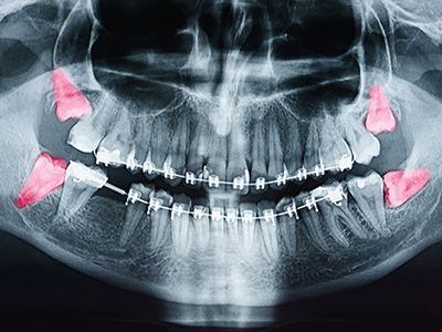
Imaging plays a central role in modern assessment. Standard panoramic X-rays give a broad view of the jaws, while cone-beam computed tomography (CBCT) or similar 3D imaging can clarify root shape, depth of impaction, and the relationship to critical structures such as the inferior alveolar nerve and sinus cavities. This level of detail helps clinicians anticipate potential complications and plan a precise, conservative approach.
Ultimately, the classification and diagnosis process is about understanding risk and choosing the timing and method of care that will result in the best long-term outcome for the patient. Clear communication about findings, options, and anticipated recovery helps patients participate in decisions about their oral health.
Extraction is typically recommended when a wisdom tooth causes symptoms or demonstrable harm to surrounding tissues. Common indications include ongoing pain, recurrent infection (pericoronitis), extensive decay that cannot be restored, cyst formation, and damage to adjacent teeth. In some cases, impacted third molars contribute to or worsen gum disease in the back of the mouth.
Decisions about removal also consider future risk. Teeth that are partially erupted or trapped at difficult angles may be more likely to develop problems over time, so proactive removal can be appropriate for younger adults who are otherwise healthy. Conversely, fully erupted, well-aligned wisdom teeth that are fully accessible for hygiene and free from disease can often be retained under regular monitoring.
Treatment planning weighs the immediate benefits of relieving symptoms against the short-term risks inherent in any surgical procedure. Factors such as the depth of impaction, proximity to nerves and sinuses, the patient’s health history, and smoking status are all part of the clinical calculus. Conservative case selection and thorough preoperative assessment reduce the chance of preventable complications.
Where there is uncertainty, the team will review alternative approaches and the likely outcomes of watchful waiting versus extraction. This collaborative discussion helps patients understand not just what will be done, but why it’s the most appropriate course for their specific clinical situation.
When extraction is indicated, the procedure is guided by careful preparation to ensure safety and comfort. Before scheduling, patients undergo a review of medical history and current medications, and imaging is reviewed to confirm the surgical plan. In many cases, cone-beam imaging or panoramic radiographs are used to map the tooth’s position relative to surrounding anatomy.
Procedures are performed under appropriate anesthesia and, when necessary, sedation. Options range from local anesthesia for straightforward cases to deeper sedation or general anesthesia for more complex situations or for patients who prefer it. The clinician will discuss the recommended level of anesthesia and answer questions about what to expect before, during, and after the procedure.
Technically, extraction of impacted third molars may require removing a small amount of bone to free the tooth and, in some cases, sectioning the tooth into pieces for gentle removal. Throughout the process, steps are taken to minimize trauma to the surrounding tissues. The clinician will explain any particular concerns—such as proximity to nerves or sinuses—and the measures used to protect those structures.
Following removal, the surgeon places gauze to control bleeding and provides specific postoperative instructions. Pain management strategies, the potential need for antibiotics, and the schedule for follow-up visits are discussed in advance so patients leave with a clear, realistic plan for recovery.
Healing after wisdom tooth extraction typically progresses quickly, but the first 48–72 hours are pivotal. Patients can expect some swelling, mild to moderate discomfort, and minimal bleeding during this time. Careful adherence to postoperative instructions—such as resting, using cold compresses early on, and avoiding vigorous rinsing or suction—helps reduce bleeding and swelling and supports clot stability at the extraction sites.
Oral hygiene remains important during recovery. Gentle rinsing with a saline solution after the initial 24 hours and careful brushing away from the extraction area minimize the risk of infection while preserving the healing clot. Dietary adjustments to soft, nutrient-dense foods for several days help maintain comfort without placing stress on the surgical sites.
Most patients return to normal activities within a few days to a week, though complete bone and soft tissue remodeling continues for several months. Any signs of prolonged or worsening pain, persistent swelling, fever, or unusual drainage should prompt a prompt call to the office. Follow-up appointments allow the clinician to confirm proper healing and address any concerns.
With appropriate technique, planning, and aftercare, the vast majority of patients experience a smooth recovery and restore comfort and function to the back of the mouth. The practice emphasizes evidence-based protocols to promote healing while minimizing the chance of complications.
We encourage patients to discuss their individual situation with our team so that treatment decisions reflect both current clinical findings and personal preferences. At Granby Dental Center, our goal is to provide clear information, thoughtful planning, and gentle, precise care. If you have questions about wisdom teeth, recovery expectations, or whether extraction is right for you, please contact us for more information.


Wisdom teeth, also called third molars, are the final permanent teeth to emerge, typically in the late teens to mid-twenties. For some people these teeth come in straight and function normally, but for many others limited jaw space or unusual angulation causes eruption problems. When a tooth cannot fully emerge or is difficult to clean, it can increase the risk of decay and gum inflammation.
Understanding how and when third molars develop helps patients and clinicians decide whether to monitor or intervene. Early evaluation allows for less invasive options when problems are identified. The goal is to preserve healthy tissue while preventing issues that could affect long-term oral health.
Impacted wisdom teeth are described by what obstructs their eruption and by their orientation. Clinicians commonly distinguish soft tissue impactions, where the tooth is covered by gum only, from bony impactions, where part or all of the tooth is encased in jawbone, and they note whether the tooth is vertical, angled or horizontal.
Classification informs the treatment approach because depth and orientation affect surgical access and the risk profile. Accurate categorization helps the team plan a conservative procedure and anticipate any special techniques that may be needed.
Common signs that a wisdom tooth may be problematic include localized pain, swelling, persistent bad breath or an unpleasant taste, and recurrent infections around the back tooth. Some patients experience difficulty opening the mouth fully (trismus) or discomfort that radiates to the jaw or ear. Repeated symptoms of inflammation or infection are clear signals to seek evaluation.
Not all problematic teeth produce obvious symptoms, so routine dental exams and appropriate imaging during adolescence or early adulthood can reveal risk factors before acute problems occur. Early detection often simplifies care and reduces the chance of complications from delayed treatment.
Diagnosis starts with a thorough medical and dental history and a clinical exam of the mouth to identify signs of infection, decay, or pressure on adjacent teeth. Symptoms guide the extent of evaluation, and even asymptomatic patients may be imaged if routine exams suggest potential future problems. Listening to the patient’s concerns is an important part of forming an individualized plan.
Imaging is essential for precise assessment: panoramic X-rays show overall jaw relationships, while cone-beam computed tomography (CBCT) or other 3D imaging clarifies root anatomy, depth of impaction and proximity to structures like the inferior alveolar nerve or sinuses. This information allows clinicians to estimate risk and choose the safest, most effective approach.
Extraction is usually recommended when a wisdom tooth causes symptoms or demonstrable harm, such as persistent pain, recurrent infections (pericoronitis), extensive decay that cannot be restored, cyst formation, or damage to adjacent teeth. Clinicians also consider long-term risk: partially erupted or severely angled teeth are more likely to cause trouble over time. The decision balances immediate benefits against the short-term risks of surgery.
Conversely, fully erupted, well-aligned wisdom teeth that can be cleaned and show no signs of disease are often monitored with regular exams and imaging. Treatment planning is individualized and takes into account the patient’s age, general health, diagnostic findings and personal preferences to arrive at the best option.
Before the procedure a clinician reviews your medical history, current medications and imaging to confirm the surgical plan and level of anesthesia needed. Many cases are managed under local anesthesia, while more complex removals may require deeper sedation or general anesthesia; the team will explain recommended options and safety measures ahead of time. Preoperative planning is aimed at minimizing surprises and ensuring patient comfort.
During surgery the provider may remove a small amount of bone and, if necessary, section the tooth into pieces to allow gentle removal while protecting surrounding structures. After extraction gauze is placed to control bleeding and you will receive clear postoperative instructions covering pain management, activity restrictions and follow-up care.
Available options range from local anesthesia, which numbs the area, to nitrous oxide, oral or intravenous sedation, and general anesthesia for deeper levels of unconsciousness. The choice depends on the complexity of the extraction, your medical history, anxiety level and the clinician’s recommendations. Each option has specific preparation and monitoring requirements to ensure safety.
Discussing your complete health history, current medications and prior experiences with anesthesia helps the team select the most appropriate approach. If sedation or general anesthesia is planned, you will receive instructions about fasting and arranging transportation home after the procedure.
Like any surgical procedure, removal of third molars carries possible risks, including infection, dry socket, bleeding, nerve irritation or injury, and, in rare cases, communication with the sinus. The likelihood of these events varies with impaction depth, root anatomy and proximity to nerves or sinuses. Understanding these possibilities is part of making an informed decision.
Risk is reduced through careful preoperative assessment, detailed imaging when indicated, conservative surgical technique and adherence to evidence-based protocols. Clear communication about potential complications, realistic expectations for recovery and close postoperative follow-up further lowers the chance of preventable problems.
Preparation begins with a review of your medical history and all medications or supplements you take, since some may affect bleeding or anesthesia choices. Follow any fasting or medication instructions provided by the clinician, and arrange for a responsible adult to drive you home if you will receive sedation or general anesthesia. Planning a few days of reduced activity and having soft foods on hand will make recovery easier.
Quit smoking if possible in the days before and after surgery because tobacco use impairs healing and increases the risk of complications. Ask the office for any specific instructions about medications, hygiene and what to bring on the day of the procedure so you arrive ready and well-informed.
Recovery typically involves some swelling and mild to moderate discomfort, with the first 48 to 72 hours being the most important for clot stability and reduction of swelling. Rest, short bursts of icing during the first 24 hours and following activity restrictions help control swelling and bleeding. Avoid vigorous rinsing, spitting or using straws initially to protect the healing clot.
After the first day gentle saline rinses and careful brushing away from the extraction sites support hygiene without disrupting healing. Maintain a soft, nutritious diet and contact the office promptly for worsening pain, fever, prolonged bleeding or unusual drainage. The team at Granby Dental Center is available to answer questions and provide follow-up care to ensure a smooth recovery.

Scheduling your next visit to Granby Dental Center is quick and hassle-free. Whether you have a specific question about our services or just need to easily book a routine cleaning, our professional staff is here to provide clear answers and simple solutions.
We’ve made it easier than ever to get in touch: give us a call or use our quick online form. Don't put your oral health on the back burner, connect with us today and let us handle the details while you focus on your smile.