Traditional dental impressions often mean trays, impression material and the very real possibility of retakes. Digital scanning with the iTero® system replaces that workflow with a cleaner, more predictable process that reduces chair time and patient discomfort. For many procedures, this shift improves communication between patient and clinician by translating what’s happening in the mouth into an instantly viewable digital model.
Beyond comfort, digital impressions change how clinicians plan treatment. High-resolution scans capture details that are difficult to preserve with physical molds, such as soft-tissue contours and undercuts. Those data become a precise reference for designing restorations, guiding implant placement or planning orthodontic movement.
Adoption of digital scanning is becoming a standard of care in modern practice because it supports better clinical decisions and fewer surprises during laboratory fabrication. At the same time, patients benefit from a more transparent, understandable process—seeing their smile in three dimensions helps set realistic expectations and fosters collaborative treatment planning.
Patients often ask whether a digital scan feels different from a standard impression. The short answer is yes—in a positive way. The iTero wand is compact and maneuverable, allowing the clinician to capture complete arches and bite relationships without triggering the gag reflex or using heavy impression materials. Most scans take only a few minutes depending on the scope of the case.
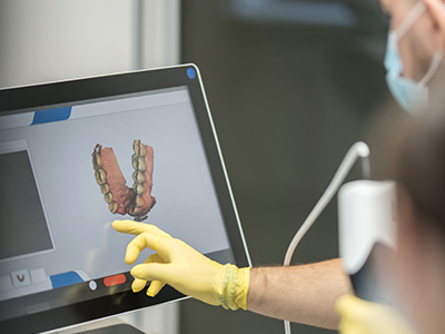
During the appointment, you’ll be able to watch the scan develop on a monitor. This live view is not only educational but also practical: it helps the clinician verify that all necessary surfaces are recorded before the patient leaves the chair. If an area needs a quick supplemental pass, that can be done immediately rather than waiting for a lab to report an inadequate impression.
The process is radiation-free and noninvasive, making it suitable for a wide range of patients, including those who have difficulty with conventional impression techniques. Because the scan is digital from the outset, it’s straightforward to store, share and reference later—useful for monitoring changes or coordinating care with specialists and laboratory partners.
The iTero scanner uses structured light and sophisticated optical software to build a high-accuracy 3D model of teeth and soft tissues. As the wand moves across the dentition, frames are captured and stitched together by the system’s processing engine to form a continuous, navigable model. This method avoids the distortions that can occur when physical impressions are poured and transported.
Real-time processing allows the clinician to inspect margin detail, occlusion and interproximal contacts immediately. That feedback loop reduces the risk of remakes and improves the fit of restorations such as crowns, bridges and onlays. For orthodontic cases, the same scan can be used to simulate tooth movement and evaluate treatment options without additional appointments.
Because the data are digital, they integrate seamlessly with CAD/CAM workflows and modern dental laboratories. This connectivity shortens turnaround times for prosthetics and aligner-based treatments while preserving the fidelity of the impression from scan to final appliance.
Precision matters when fabricating restorations and appliances. Digital impressions produced by the iTero system capture fine anatomic details that help labs create better-fitting crowns, veneers and implant abutments. Tight margins and accurate occlusal contacts translate into restorations that require less chairside adjustment and provide more reliable long-term performance.
In orthodontics, digital scans streamline the diagnostic process and the production of clear aligners. The ability to produce a treatment simulation based on the patient’s actual anatomy improves case acceptance and makes the progression of care more predictable. For multidisciplinary cases, a shared digital model becomes a single source of truth for all team members involved.
Additionally, the traceability of digital files provides a useful medico-legal and clinical record. Scans can be archived indefinitely and revisited to assess wear, recession, or tooth movement over time—information that’s valuable for preventive care and ongoing maintenance.
Patient experience is a major reason many practices transition to digital impressions. Eliminating bulky trays and impression paste reduces anxiety for patients with sensitive gag reflexes or strong aversions to traditional impressions. Shorter, more comfortable appointments also contribute to higher satisfaction and a smoother workflow for the dental team.
From an operational perspective, digital impressions reduce the number of consumables and the logistical overhead associated with shipping and storing physical models. Files can be transmitted instantly to a laboratory or to an in-house milling unit, accelerating turnaround without compromising accuracy.
Safety is another consideration: the iTero scanning process is noninvasive and free of ionizing radiation. For patients who require frequent monitoring—such as orthodontic patients or those undergoing phased restorative treatment—having a safe, repeatable method of documentation is highly advantageous.
Integration is where digital impressions often deliver their greatest value. A single iTero scan can be used across diagnostic, restorative and orthodontic workflows, allowing teams to coordinate care more efficiently. For example, the same dataset can inform a diagnostic wax-up, guide surgical guides for implants, or be used to design aesthetic veneers.
Lab communication becomes more streamlined when technicians receive standardized digital files. Fewer interpretation steps and less manual handling reduce opportunities for error, and labs can provide faster, more consistent results. For practices that use in-office milling or printing, digital scans can enable same-day restorations for selected cases, improving convenience for patients.
At the practice level, adopting a reliable scanner supports longitudinal care. Clinicians can compare scans over time to track outcomes, measure tissue changes, and document the progression of treatment. This continuity elevates the quality of care and reinforces clinical decision-making with objective data.
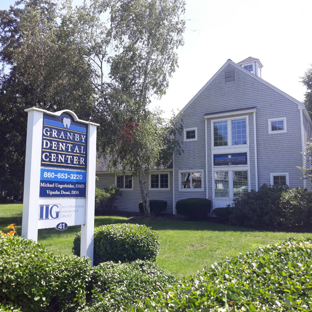
Granby Dental Center embraces digital technologies like the iTero® system to enhance diagnostic clarity and patient comfort while maintaining a focus on predictable, evidence-based care.
To learn more about how digital impressions may benefit your treatment, please contact us for additional information or to schedule a consultation.

The iTero® digital impression system is an intraoral scanner that uses structured light and advanced optical software to create high-resolution three-dimensional models of teeth and soft tissues. As the wand moves across the dentition, captured frames are stitched together by the system to form a continuous, navigable model that clinicians can inspect in real time. This digital approach replaces physical trays and impression materials for many procedures while preserving fine anatomic detail.
Captured scans serve as an accurate reference for designing restorations, planning implant placement and simulating orthodontic movement. Because the data are digital from the outset, they integrate directly with CAD/CAM workflows and modern dental laboratories. The result is a predictable digital record that clinicians can use for diagnostics, planning and communication with treatment partners.
Digital scans eliminate the need for bulky trays and impression paste, reducing the risk of distortion associated with material handling and shipping. The scanner records soft-tissue contours, undercuts and occlusal relationships with high fidelity, avoiding many of the dimensional changes that can occur when physical impressions are poured and stored. Real-time visualization also allows clinicians to confirm complete capture before the patient leaves the chair.
Because the file is digital, it can be transmitted instantly to a laboratory or used in an in-office CAD/CAM system, shortening turnaround time and reducing the likelihood of remakes. The streamlined workflow decreases logistical overhead such as shipping and storage of physical models. Overall, digital scanning tends to produce a more consistent and transparent process for both clinicians and patients.
During an iTero® scan the clinician will move a compact, handheld wand around the teeth while the scanner captures a series of images that are processed into a 3D model. Most complete-arch scans take only a few minutes depending on the scope of the case, and patients can often watch the progress on a chairside monitor. The process is noninvasive and designed to minimize discomfort, making it easier for patients who have a sensitive gag reflex or difficulty with conventional impressions.
Clinicians use the live view to verify that all necessary surfaces are recorded and to perform quick supplemental passes if needed, which reduces the chance of returning for a retake. At the office of Granby Dental Center, this immediate feedback helps explain findings and set realistic expectations before treatment proceeds. Because the scan is digital, it is also straightforward to store and share with specialists or laboratories when coordinating care.
Yes, the iTero® scanning process is noninvasive and free of ionizing radiation, making it safe for a broad range of patients including children, adults and those who require frequent monitoring. There is no exposure to x-rays during the scanning itself, and the procedure generally requires no special preparation. Because it avoids impression materials, patients with sensitivities to paste are less likely to experience adverse reactions during the appointment.
Clinicians will still evaluate individual limitations, such as restricted mouth opening or severe soft-tissue inflammation, and adapt their approach as needed to ensure a complete capture. For patients who need serial records, the safety and repeatability of digital scans make them an efficient option for monitoring progress over time. Overall, the technique is considered low risk and well suited for routine diagnostic and treatment documentation.
In restorative dentistry iTero® scans capture margin detail, occlusal contacts and interproximal anatomy that laboratories use to design precise crowns, veneers and implant abutments. High-resolution digital impressions reduce the need for chairside adjustments by improving the initial fit of laboratory-fabricated restorations. This precision helps streamline the delivery process and supports long-term restoration performance.
Because the data integrate with CAD/CAM systems, laboratories and in-office milling units can use the same dataset to fabricate restorations with fewer interpretation steps. The standardized digital workflow reduces opportunities for error related to physical model handling and transportation. Clinicians can also archive scans as a reference for future restorative needs or for verifying pre- and post-treatment conditions.
iTero® scans provide accurate digital models that are used to plan orthodontic treatment, create clear aligners and simulate anticipated tooth movement. Treatment simulations generated from the patient’s actual anatomy improve case visualization and help patients understand the planned progression of care. The same scan used for diagnostics can be sent to aligner manufacturers or used to track incremental changes over time.
Serial scanning allows clinicians to compare models across appointments, monitor progress and document outcomes objectively. For multidisciplinary cases, the shared digital model becomes a single source of truth for all team members involved in treatment. This interoperability enhances coordination between orthodontists, restorative clinicians and laboratory partners.
Digital scanning produces standardized files that can be transmitted instantly to laboratories and specialists, reducing turnaround time and limiting the potential for interpretation errors. Technicians receive high-fidelity data that fit directly into CAD/CAM workflows, which simplifies the design and fabrication process. The clarity of the digital model also enables more precise instructions and fewer follow-up questions that can delay production.
For complex or multidisciplinary cases, clinicians can share the dataset along with annotations to coordinate surgical guides, prosthetic designs or orthodontic plans. This shared digital environment promotes consistency across all contributors and supports efficient decision-making. The result is a smoother lab-to-chair workflow and improved predictability of clinical outcomes.
Yes, digital scans can be archived indefinitely and serve as reliable records for long-term treatment planning and monitoring. Stored models allow clinicians to compare anatomy over time, evaluate wear patterns, track recession or tooth movement, and document outcomes for preventive care. The traceability of digital files also provides useful clinical and medico-legal documentation when needed.
Because the files are digital, they are easy to retrieve and share when coordinating phased treatment or consulting with specialists. This accessibility supports continuity of care by enabling clinicians to revisit baseline scans during subsequent visits. The ability to reference past scans reduces redundant data collection and enhances informed clinical decision-making.
In many cases, iTero® scans shorten the time required for obtaining impressions by removing the steps associated with tray seating, material setting and physical model pouring. Real-time verification allows clinicians to address incomplete captures immediately, which reduces the frequency of remakes and follow-up appointments. This efficiency can lead to smoother chairside workflow on scanning days.
Operationally, digital impressions also reduce consumable usage and logistical demands related to shipping and storing physical models. When combined with in-office milling or reliable lab partners, scans can enable faster turnaround for restorations and aligner-based treatments. The streamlined process improves team productivity while maintaining clinical accuracy.
Granby Dental Center adopts the iTero® system to enhance diagnostic clarity, improve patient comfort and support predictable, evidence-based care. The scanner’s ability to capture detailed anatomy and provide immediate visualization helps clinicians make informed treatment decisions and communicate options clearly to patients. Using a digital workflow also supports better coordination with laboratories and specialists across restorative and orthodontic cases.
By integrating digital impressions into routine practice, the team reduces the likelihood of remakes, shortens turnaround times and preserves a detailed archive for longitudinal care. This approach aligns with the practice’s goal of delivering efficient, transparent and well-documented treatment plans. Patients benefit from a more comfortable experience and clinicians benefit from objective data that inform treatment at every stage.
Scheduling your next visit to Granby Dental Center is quick and hassle-free. Whether you have a specific question about our services or just need to easily book a routine cleaning, our professional staff is here to provide clear answers and simple solutions.
We’ve made it easier than ever to get in touch: give us a call or use our quick online form. Don't put your oral health on the back burner, connect with us today and let us handle the details while you focus on your smile.