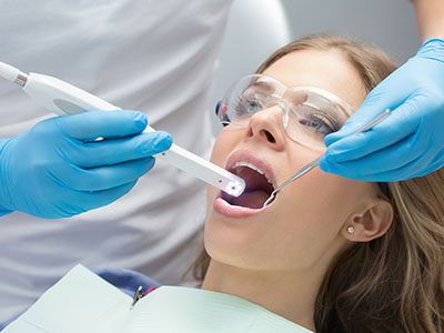
An intraoral camera is a compact, pen-sized imaging tool designed to capture clear, full-color photographs of the teeth, gums, and other soft tissues inside the mouth. Unlike external photography, these cameras are optimized for the confined spaces of the oral cavity, producing high-resolution images in real time. The resulting photos appear on a chairside monitor, giving both clinician and patient an immediate, magnified view of oral anatomy and areas that may warrant attention.
Because the device is small and maneuverable, it can reach angles and surfaces that are difficult to see with the naked eye or even with traditional mouth mirrors. It often uses LED illumination and macro lenses to highlight detail and color contrast, which helps clinicians distinguish surface texture, early demineralization, and subtle tissue changes. This level of visual detail supports more precise observation than routine visual inspection alone.
Captured images can be paused, enlarged, and reviewed frame-by-frame, allowing the dental team to document findings accurately and consistently. The live feed and still images also integrate with digital patient records, creating a visual baseline that can be referenced during follow-up visits. In practical terms, the intraoral camera transforms fleeting visual information into permanent, shareable data that supports diagnosis and continuity of care.
One of the most immediate benefits of intraoral imaging is how it improves communication. When patients can see a clear, magnified image of their own teeth and gums, explanations about conditions and recommended treatments become more concrete. Visual evidence helps bridge the gap between technical clinical language and a patient’s understanding, fostering informed decision-making and reducing confusion about oral health status.
Using the intraoral camera as a teaching tool also encourages collaborative care. Dentists can point out early enamel changes, plaque accumulation, or restorative margins directly on the screen, demonstrating why a particular approach is recommended. This transparency builds trust and helps patients prioritize oral hygiene or accept preventive interventions earlier, when they are most effective.
For patients who may be anxious or skeptical, seeing real-time images normalizes the clinical environment and demystifies procedures. Visuals can highlight improvement over time, showing the positive effects of home care or professional treatment in a way that words alone cannot. In short, intraoral photography turns abstract diagnoses into visible, actionable information.
At Granby Dental Center, we employ intraoral imaging routinely to involve patients in their care and to ensure that treatment choices are clearly explained and well documented for future reference.
Intraoral cameras play a key role in early detection of dental issues. High-resolution images can reveal enamel changes, hairline fractures, and restorative defects that are easily missed during a cursory exam. Spotting these concerns sooner allows for conservative interventions that preserve more natural tooth structure and prevent more extensive problems down the line.
Beyond detecting decay and cracks, these images support diagnosis of soft-tissue conditions such as inflammation, mucosal lesions, and abnormal pigmentation. When paired with a thorough clinical assessment, intraoral photographs contribute to a more complete diagnostic picture and can prompt timely referrals to specialists when appropriate.
The technology also enhances documentation accuracy. When a clinician documents a finding with a clear image, it reduces ambiguity in records and supports consistent monitoring. For complex restorative cases, precise intraoral photos assist in communication with dental laboratories and specialists, ensuring that color, contour, and margin details are captured and conveyed effectively.
Finally, intraoral imaging complements other diagnostic tools—such as radiographs—by providing surface-level visualization. Together, these modalities create a layered approach to diagnosis that improves clinical confidence and treatment outcomes.
Images captured by intraoral cameras are saved directly into the patient’s digital chart, becoming part of a permanent clinical record. This visual history is invaluable for tracking the progression of conditions, comparing pre- and post-treatment results, and documenting the rationale behind clinical decisions. Clear photographic records make longitudinal care more transparent and reduce the risk of miscommunication across multiple visits or providers.
When treatment planning, these images help the dental team create more accurate, customized plans. Photographs can be annotated to mark areas of concern, included in case notes, and shared with labs or specialists to convey exact requirements. Visual documentation is particularly helpful in coordinating multidisciplinary care, such as restorative work that relies on both dentist and lab technicians.
Because images are archived alongside chart notes, they also support quality assurance and clinical review. The ability to review previous images side-by-side with new ones streamlines follow-up assessments and helps clinicians evaluate the effectiveness of preventive measures or treatments over time.
An intraoral camera exam is quick, comfortable, and noninvasive. During a routine visit, your dental professional will position the device inside the mouth to photograph specific teeth or soft-tissue areas. The process typically takes only a few minutes and is painless; the camera’s small profile makes it easy to maneuver around the arches without causing discomfort.
As images appear on the monitor, your clinician will explain what you’re seeing, point out areas that need attention, and discuss possible next steps. This real-time review allows you to ask questions and to see the same visual evidence the clinician is using to make recommendations. If additional photos are needed—such as close-ups of a particular restoration—they can be taken and saved immediately.
After the exam, the selected images are stored in your record for future reference and for inclusion in treatment plans when necessary. If a referral or laboratory communication is required, these images can be included to ensure continuity of care and precise instructions from one provider to another.
The office of Granby Dental Center uses intraoral imaging as a standard part of exams to support clear communication and to help patients participate in care decisions with confidence.
In summary, intraoral cameras are a practical, patient-friendly technology that improves visualization, communication, and clinical documentation. They help clinicians detect issues earlier, explain findings more clearly, and plan treatment with greater precision. If you’d like to learn more about how intraoral imaging is used during exams or how it might benefit your dental care, please contact us for more information.


An intraoral camera is a small, pen-sized imaging device designed to photograph the teeth, gums and other soft tissues inside the mouth. It captures high-resolution, full-color images using a macro lens and LED illumination to reveal fine surface detail that can be difficult to see with the naked eye. Images are displayed in real time on a chairside monitor, allowing clinicians and patients to review the oral anatomy together. The device is maneuverable enough to reach tight angles and document areas of interest for diagnosis and record keeping.
The live feed can be paused, enlarged and reviewed frame-by-frame so clinicians can document findings consistently and compare images over time. Still images are commonly saved directly into the patient’s digital chart to create a visual baseline for future exams. Because intraoral cameras translate fleeting observations into permanent images, they improve the accuracy of clinical documentation. They also enable clearer communication with laboratories and specialists when detailed surface information is needed.
Intraoral imaging enhances the clinician’s ability to detect early enamel changes, hairline fractures, restorative defects and subtle tissue abnormalities that routine visual inspection might miss. High-resolution photographs make surface texture, color differences and marginal discrepancies more apparent, which supports earlier and more conservative intervention when appropriate. The images help clinicians identify issues at an earlier stage, often before they become more extensive. Early detection can preserve more natural tooth structure and simplify treatment options.
These photographs are also valuable for evaluating soft-tissue concerns such as localized inflammation, mucosal lesions or pigmentary changes that warrant monitoring or referral. When combined with a full clinical exam and other diagnostic tools, intraoral images contribute to a more complete diagnostic picture. Clinicians can use the documented images to decide when to escalate care or involve specialists. At the office of Granby Dental Center, clinicians integrate intraoral imaging into routine exams to support timely, evidence-based decisions.
An intraoral camera exam is quick, noninvasive and generally well tolerated by most patients. The device has a small profile that can be maneuvered around the dental arches with minimal discomfort, and captures images within a matter of minutes. Because it is not a radiographic tool, there is no exposure to ionizing radiation when using the camera. Patients can watch the images on the monitor as they are taken, which often reduces anxiety by making the examination more transparent.
Clinics follow infection-control protocols such as disposable sleeves or barrier covers and routine device cleaning to maintain patient safety. The LED light sources used in modern intraoral cameras are designed for short-duration clinical use and are considered safe for inspection purposes. If a patient has specific concerns about comfort or oral sensitivity, the clinician can adjust technique or spacing to minimize any discomfort. Overall, the procedure is a low-risk adjunct to a standard dental exam.
Intraoral photographs are integrated into the patient’s digital record and used to inform individualized treatment plans. Clinicians can annotate images to mark areas of concern, include them in case notes and use them to explain recommended procedures to patients. This visual documentation helps the dental team create more accurate and predictable restorative plans by preserving details about color, contour and margin relationships. Images also serve as a reference for monitoring progress before, during and after treatment.
Because images can be shared securely with dental laboratories and specialists, they improve the clarity of instructions when collaborative care is required. Accurate visuals reduce ambiguity and help technicians reproduce desired outcomes more consistently. The ability to show patients the same images used by the clinical team fosters shared decision-making and makes treatment goals easier to understand. Visual records therefore streamline both clinical workflow and multidisciplinary coordination.
Intraoral cameras and radiographs provide complementary information rather than serving as direct substitutes for one another. Cameras excel at surface-level visualization, revealing enamel defects, cracks, restorative margins and soft-tissue changes that are visible externally. X-rays, by contrast, provide essential information about internal tooth structure, root anatomy and underlying bone that cameras cannot capture. Relying on both modalities gives clinicians a layered diagnostic approach that improves overall detection and treatment planning.
For example, a hairline fracture or a poorly fitting restoration may be obvious with intraoral imaging but invisible on a radiograph, while early interproximal decay or bone loss may require radiographic imaging for confirmation. Clinicians use the strengths of each tool together to form a complete assessment. This combined use helps avoid missed diagnoses and supports more targeted, evidence-based care.
Images captured by intraoral cameras are typically saved directly into the patient’s electronic chart as part of the official clinical record. Dental offices use secure practice management systems that include measures such as user access controls, audit logs and data encryption to protect patient information. Proper account permissions limit who can view or modify records, and routine backups preserve images for long-term comparison. The digital format makes it simple to retrieve and review previous images alongside new ones during follow-up visits.
When images are shared with outside providers or laboratories, clinicians follow privacy and consent protocols to ensure data is transmitted securely and only to authorized recipients. Visual records support continuity of care by reducing ambiguity in referrals and by allowing receiving clinicians to examine documented findings remotely. Retained images also facilitate quality assurance and help clinicians evaluate the effectiveness of treatments over time.
Seeing a clear, magnified image of your own teeth and gums bridges the gap between clinical language and personal understanding. Visual evidence makes it easier to comprehend the nature and extent of findings, which supports informed consent and shared decision-making. When patients can view the same images the clinician is using, questions become more specific and discussions about options are more productive. This transparency reduces uncertainty and helps patients prioritize interventions that align with their goals.
Visual documentation also motivates preventive behavior by showing areas of plaque accumulation or early enamel change that can respond to improved home care. Over time, successive images illustrate treatment progress or the benefits of preventive measures, reinforcing adherence to recommended routines. In short, intraoral imaging converts abstract descriptions into concrete visuals that empower patients to participate actively in their care.
In most cases, intraoral imaging adds only a few minutes to a routine dental visit because modern cameras capture high-quality images rapidly and integrate smoothly into the exam workflow. Clinicians typically take a set of standard photographs and then capture any additional close-ups that are needed for specific concerns. The time spent reviewing images with patients is usually brief and focused, and it often reduces follow-up questions by clarifying findings during the appointment. When complex documentation is required for restorations or referrals, capturing and annotating a few extra images may extend the visit slightly.
Because images are saved directly to the digital chart, the documentation step is efficient and reduces the need for repetitive exams later. The straightforward nature of the process means most patients experience little or no disruption to the overall appointment time. Any additional minutes contribute to clearer communication and more precise treatment planning.
Intraoral images capture critical surface-level details such as shade, contour and margin relationships that are important for aesthetic and functional restorative work. Photographs can be annotated and included with laboratory prescriptions to convey precise visual instructions that complement written notes and impressions. Clear visuals reduce ambiguity about color matching and margin placement, which helps technicians fabricate restorations that better meet the clinician’s and patient’s expectations. Accurate photographic communication can lessen the need for adjustments and improve overall case predictability.
Images are also useful when coordinating complex multidisciplinary cases or when a specialist needs to evaluate a restoration remotely. By providing high-quality visuals, the dental team can discuss details more efficiently and ensure that laboratory and clinical objectives are aligned. Photographic documentation therefore strengthens collaboration across the restorative workflow and supports more consistent outcomes.
An intraoral camera exam is typically brief and begins with the clinician positioning the device to capture images of specific teeth or soft-tissue areas. As images appear on the chairside monitor, the clinician will explain what is being shown and point out any findings that require attention. You will have the opportunity to ask questions in real time and to view the same visual evidence used to make recommendations. If closer documentation is needed, additional close-up photos may be taken and saved immediately.
After the exam, selected images are archived in the patient’s digital record for future reference and for inclusion in treatment planning or referral communications. The process is a routine part of many modern dental exams and is intended to improve understanding, documentation and continuity of care. If you have questions about how intraoral imaging will be used in your care, the office of Granby Dental Center can explain the protocol during your visit.
Scheduling your next visit to Granby Dental Center is quick and hassle-free. Whether you have a specific question about our services or just need to easily book a routine cleaning, our professional staff is here to provide clear answers and simple solutions.
We’ve made it easier than ever to get in touch: give us a call or use our quick online form. Don't put your oral health on the back burner, connect with us today and let us handle the details while you focus on your smile.