Digital radiography replaces traditional film with electronic sensors that capture X-ray images and send them directly to a computer. Instead of developing film in a darkroom, the sensor produces a high-resolution image within seconds, allowing clinicians to see the result immediately. This quick turnaround helps streamline appointments and supports more focused conversations between you and your provider.
Because images are captured as digital files, they can be enhanced, magnified, or adjusted to highlight important details without retaking the image. That capability improves diagnostic clarity for subtle problems that might be hard to see on conventional film. The result is a more thorough evaluation that supports accurate treatment planning while minimizing the need for repeat exposures.
Digital radiography also simplifies recordkeeping. Files are stored within the practice’s secure electronic charting system, where they can be compared with past images to track changes over time. This continuity of care helps dentists and hygienists measure treatment progress and make decisions rooted in a complete view of a patient’s oral health history.
At the same time, digital systems are designed to meet modern safety and privacy standards. Sensors and software are regularly maintained and updated, and images are stored according to professional guidelines, ensuring both clinical reliability and patient confidentiality.
For patients, one of the most noticeable advantages of digital radiography is speed. Because images appear immediately on a monitor, the clinician can review findings with you during the same visit and explain recommended next steps without delay. This instant feedback reduces anxiety and helps you make informed choices about your care while you’re in the chair.
Digital sensors are typically smaller and more flexible than traditional film packets, which can make the imaging process more comfortable for many patients. Shorter exposure times are common with digital systems, contributing to an overall reduction in radiation dose compared with older film techniques. Those improvements are part of routine efforts to make diagnostic imaging as safe as possible.
Another patient benefit is the environmental advantage: because digital radiography eliminates the need for chemical developers and paper-based storage, it reduces waste associated with conventional film processing. That lower environmental impact is an additional way modern practices can provide responsible, contemporary care.
Finally, digital images make it easier for clinicians to include you in treatment discussions. With on-screen visualization, you can see the same images your provider uses, making explanations clearer and helping you understand the rationale for recommended procedures.
Digital radiographs are a powerful diagnostic tool because software allows clinicians to adjust contrast, zoom in on areas of interest, and apply filters that reveal details difficult to detect on film. Those enhancements can expose early signs of decay, changes in bone levels, or issues around restorations that would otherwise be less obvious.
Because the images are electronic, they can be shared quickly and securely with specialists when a referral or consultation is required. This seamless collaboration accelerates multidisciplinary care—whether coordinating with an oral surgeon, periodontist, or endodontist—and helps ensure everyone involved has the same clear visual information to guide treatment.
Digital files also support better long-term monitoring. Side-by-side comparisons of current and past images allow clinicians to track healing, evaluate the stability of restorations, and document progressive conditions in a way that’s objective and measurable. That historical perspective contributes to safer, more predictable treatment outcomes.
Integration with diagnostic tools and software further enhances clinical decision-making. From treatment simulation to pre-surgical planning, digital radiography plays an important role in delivering precise, evidence-based care.
Digital radiography is not an isolated technology; it connects with electronic health records, treatment planning systems, and patient education tools to form a cohesive clinical workflow. When images are linked to a patient’s chart, notes, and treatment history, clinicians can assemble a holistic view of oral health without sifting through paper files.
That integration supports a proactive approach to care. For example, images can be annotated during a consultation, included in treatment estimates, or used to demonstrate the expected outcomes of various options. Visual aids help patients understand complex procedures, which often leads to better-informed consent and higher satisfaction with care decisions.
In surgical or restorative cases, digital imaging contributes to precision. Clear radiographs guide implant placement, endodontic treatment, and the assessment of bone structure and tissue margins. The ability to plan with accurate visual references reduces uncertainty and supports predictable clinical results.
As digital tools continue to evolve, their role in diagnostics and treatment planning will expand. Practices that embrace these systems can offer a smoother patient experience and maintain clinical standards that align with contemporary dental medicine.
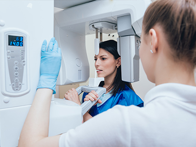
A digital radiography appointment typically follows a familiar workflow: your clinician positions a small sensor in your mouth or directs a hand-held/extraoral device, captures the image, and reviews the result on a monitor. The process is brief and designed to be as comfortable as possible while capturing diagnostically useful images.
Safety remains a priority throughout. Clinicians follow established protocols to minimize exposure, including proper sensor placement, shielding when appropriate, and limiting images to those that are clinically necessary. If you have specific concerns—such as pregnancy or sensitivity—mention them to the team so they can tailor procedures to your needs.
If you are new to the practice or receiving care coordination with another provider, digital images can often be imported or exported to support continuity of care. This flexibility reduces the chance of duplicate imaging and makes it easier for your clinician to review past records when forming a treatment plan.
After the images are taken, your provider will review them with you, explain any findings in clear terms, and outline recommended next steps. Patients appreciate the immediate clarity that digital radiography provides; seeing the images often makes explanations more tangible and helps set expectations for treatment.
Digital radiography has become a cornerstone of modern dental diagnosis due to its speed, clarity, and integration with current clinical systems. By combining improved imaging quality with streamlined workflows and enhanced patient communication, it supports safer, more informed care. If you’d like to learn more about how digital x-rays are used in our practice or what to expect during an appointment, please contact Granby Dental Center for more information.


Digital radiography replaces traditional film with electronic sensors that capture X-ray images and deliver high-resolution files to a computer within seconds. This rapid capture streamlines appointments by allowing clinicians to review images immediately and discuss findings with patients during the same visit. Because files can be enhanced and magnified, clinicians often detect subtle issues earlier than with conventional film.
Images produced by digital sensors are stored as digital records that can be compared with prior studies to monitor changes over time and support long-term treatment planning. At Granby Dental Center this capability helps the clinical team create more targeted, evidence-based care plans while reducing the need for repeat exposures. The result is a more efficient diagnostic process and clearer communication with patients about their oral health.
Digital radiography typically requires shorter exposure times than conventional film systems, which reduces the overall radiation dose for patients. Clinicians follow established safety protocols such as using proper sensor placement, shielding when appropriate, and taking only images that are clinically necessary. The practice of minimizing exposure is consistent with the ALARA principle—keeping doses as low as reasonably achievable while obtaining diagnostic-quality images.
Modern digital systems are designed with safety in mind and undergo routine checks to ensure consistent performance. If you have specific concerns, such as pregnancy or heightened sensitivity, mention them to the team so imaging can be adjusted or deferred when clinically appropriate. Staff will explain any additional precautions used to protect you and your family.
Digital images can be manipulated with software tools to adjust contrast, zoom in on areas of interest, and apply filters that reveal details difficult to see on film. These capabilities help clinicians detect early decay, assess bone levels, and evaluate the condition of restorations with greater confidence. Side-by-side comparisons of current and prior images support objective monitoring of healing and disease progression.
Because images are electronic they integrate with treatment planning systems and can be annotated to illustrate findings or surgical guides. This integration improves precision for procedures such as implant placement and endodontic therapy by providing clear visual references. The ability to simulate outcomes and review images with the patient also supports better-informed decisions and clearer consent.
A typical appointment involves positioning a small intraoral sensor or using an extraoral imaging device, capturing the necessary images, and reviewing the results on a monitor. The process is brief and designed for patient comfort, with sensors that are often smaller and more flexible than traditional film packets. Clinicians will explain the purpose of each image and answer any questions before or after capture.
Safety steps such as the use of lead aprons or thyroid collars are applied when appropriate, and technicians take care to limit exposures to what is clinically required. If you have concerns about comfort or special circumstances, speak with the dental team so they can make accommodations. After imaging, the clinician will review findings with you in plain language and outline recommended next steps.
Digital radiographs are stored as part of the practice’s electronic health record using secure systems that meet professional standards for privacy and data protection. Access to images is restricted to authorized clinical staff and is typically logged in the practice management system to preserve confidentiality. Many practices use encryption and routine backups to protect files against loss and unauthorized access.
Retention and sharing of images follow regulatory and ethical guidelines to ensure patient privacy while enabling coordinated care. When images are transmitted to other providers or specialists, secure channels and secure file formats are used to minimize risk. Patients who wish to discuss records, request copies, or understand retention policies can ask the office team for details about data handling practices.
Because digital images are electronic, they can be exported and shared quickly and securely with specialists when referrals or consultations are needed. Files are typically transmitted using secure file-transfer methods or through integrated referral platforms that protect patient information. This efficient sharing supports timely collaborative care and ensures all members of the treatment team see the same diagnostic images.
Shared images are accompanied by clinical notes and relevant patient history so specialists have the context needed to provide informed recommendations. Where appropriate, clinicians will coordinate directly with referring providers to review findings and agree on next steps. Patients should inform the office if they prefer to receive copies of their images or if they are coordinating care with an outside practice.
Digital radiography can be used safely for children and pregnant patients when clinically indicated, because sensors often allow for shorter exposures and reduced radiation compared with older film approaches. Pediatric imaging is tailored to the child’s size and developmental stage to minimize discomfort and exposure, and clinicians use the fewest images necessary for an accurate diagnosis. For pregnant patients, imaging is evaluated on a case-by-case basis and is performed only when the expected benefit outweighs any potential risk.
If you are pregnant or believe you might be pregnant, inform the dental team before imaging so they can consider alternative approaches or postpone nonurgent radiographs. Protective measures such as lead aprons and thyroid shields are used whenever appropriate to further reduce exposure. The team will discuss the clinical rationale for any recommended images and answer questions about safety.
The frequency of dental X-rays is individualized and based on each patient’s medical and dental history, risk factors, and current oral health status. Patients with active disease, a history of restorative work, or other risk factors may require imaging more frequently than those with low risk and stable oral health. Clinicians follow professional guidelines and use clinical judgment to determine the minimal schedule that still provides safe, effective care.
Routine imaging intervals are not one-size-fits-all; instead, they reflect an evidence-based balance between diagnostic benefit and exposure reduction. During checkups the clinician will evaluate whether new images are necessary and will discuss the reasoning with the patient. Personal concerns about imaging frequency can be addressed directly with the dental team so plans reflect individual preferences and clinical needs.
While digital radiographs provide excellent two-dimensional detail for many diagnostic tasks, they cannot capture three-dimensional anatomy in the way cone-beam computed tomography (CBCT) can. Small or early lesions may still be difficult to visualize depending on angulation, overlapping structures, or image artifacts. Clinicians recognize these limitations and will recommend additional imaging when three-dimensional information or higher-resolution views are necessary.
Image quality can also be affected by sensor position, patient movement, or technical settings, which is why proper technique and periodic equipment checks are important. When a radiograph is inconclusive, the care team may repeat an image with adjusted technique or use a different modality to obtain the needed diagnostic clarity. The choice of imaging is driven by the clinical question and the goal of obtaining reliable information with minimal exposure.
Digital radiography integrates with electronic health records, treatment planning tools, and patient education platforms to create a cohesive workflow that supports coordinated care. Images can be annotated, incorporated into simulations, and used to explain procedures visually during consultations, which improves patient understanding and informed decision-making. Integration also streamlines administrative tasks such as recordkeeping and referral coordination.
Because of this connectivity, digital imaging contributes directly to precision in restorative and surgical cases by providing accurate visual references for planning and execution. The practice uses these tools to align clinical documentation, imaging, and treatment steps so that outcomes are as predictable and reproducible as possible. Continued software updates and interoperability improvements expand these capabilities over time.
Reliable digital imaging depends on routine maintenance, software updates, and quality-assurance checks that verify sensor calibration and image consistency. Clinical staff are trained to follow standardized protocols for sensor handling, positioning, and infection control to preserve image quality and safety. Regular preventive maintenance helps identify technical issues early so they do not compromise diagnostic accuracy.
The office also implements periodic image quality audits and follows manufacturer recommendations for software patches and hardware servicing to maintain performance. If a patient or clinician notices recurring image artifacts or technical problems, the team will investigate and address them promptly to ensure diagnostic reliability. These processes support consistent, high-quality imaging for every patient.
Scheduling your next visit to Granby Dental Center is quick and hassle-free. Whether you have a specific question about our services or just need to easily book a routine cleaning, our professional staff is here to provide clear answers and simple solutions.
We’ve made it easier than ever to get in touch: give us a call or use our quick online form. Don't put your oral health on the back burner, connect with us today and let us handle the details while you focus on your smile.