At Granby Dental Center, we pair clinical experience with advanced imaging to give patients clearer diagnoses and more predictable treatment outcomes. Cone-beam computed tomography (CBCT) delivers three-dimensional views of dental and facial structures that traditional X-rays cannot, helping clinicians see anatomy with greater precision and confidence. Our approach is to use that information thoughtfully—combining clear imaging with careful clinical judgment to design treatments tailored to each patient's needs.
High-quality imaging is only useful when interpreted by a knowledgeable team. We prioritize concise, patient-centered explanations so you understand what the scan shows and why it matters for your care. Whether you are exploring restorative options, evaluating a source of jaw pain, or planning oral surgery, CBCT can provide the crucial anatomical detail that leads to safer, more efficient care.
Unlike flat radiographs, CBCT captures volumetric data that reveals depth, spatial relationships, and subtle structural details. This means dental professionals can evaluate bone thickness, root canal anatomy, and the proximity of critical structures such as nerves and sinuses with much greater accuracy. Those extra dimensions reduce uncertainty and help avoid surprises during treatment.
For patients, the practical benefit is a clearer plan and fewer unexpected steps. When clinicians can visualize anatomy from multiple angles, they can refine treatment choices, estimate procedural complexity more reliably, and choose techniques that minimize invasiveness. The result is a smoother clinical experience with a stronger emphasis on predictable outcomes.
CBCT also supports better communication among specialists. Detailed 3D images enable more precise referrals and collaborative planning between general dentists, oral surgeons, endodontists, and orthodontists. That interdisciplinary clarity often shortens treatment timelines and improves coordination of care.
Cone-beam CT uses a cone-shaped X-ray beam and a detector that rotates around the head to capture a series of images. These individual exposures are reconstructed into a three-dimensional dataset that can be viewed as slices or as a volumetric model. The technology is designed to focus on the area of interest, which helps limit radiation exposure compared with some medical CT protocols.
Modern CBCT systems provide the flexibility to select smaller fields of view for focused exams—reducing unnecessary exposure while still delivering the diagnostic detail clinicians need. Technicians follow industry best practices for positioning and exposure settings, and scans are performed only when clinically justified to ensure patient safety.
Because the images are digital, they can be manipulated in real time to highlight specific structures or to create measurements used for planning. This efficiency speeds up decision-making and reduces the need for multiple appointments or repeat imaging when clear information is already available.
CBCT is invaluable for implant planning because it shows both bone quantity and quality in three dimensions. Accurate assessment of ridge anatomy and the relationship to adjacent teeth or anatomical landmarks allows clinicians to select appropriate implant size and position, anticipate the need for bone grafting, and plan surgical guides when required.
Endodontics is another area where CBCT is frequently helpful. Complex root canal systems, suspected fractures, or persistent symptoms after treatment can be evaluated with more clarity using three-dimensional imaging. That additional information helps clinicians identify the true cause of a problem and choose the most effective approach.
Beyond implants and endodontics, CBCT supports airway assessment, TMJ evaluation, pathology detection, and orthodontic treatment planning. The versatility of the scan makes it a useful diagnostic adjunct across many dental specialties, providing a single dataset that can be reviewed and reinterpreted as treatment needs evolve.
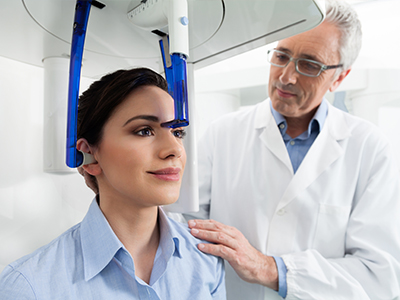
A CBCT scan is typically quick and noninvasive. Patients are positioned comfortably—either standing or seated—while the scanner rotates around the head for a matter of seconds to capture the images. There is no need for confined spaces like those associated with some medical CT units, and no sedation is required for routine scans.
Staff take steps to ensure patient comfort and safety throughout the procedure. Standard precautions such as protective shielding and careful positioning are used when appropriate, and the imaging team verifies clinical indications before proceeding. Because scans can be completed rapidly, the impact on your appointment time is minimal.
After the acquisition, the images are reviewed by the treating clinician, who will explain the findings in plain language and outline how the information informs the next steps in care. If a specialist consult is recommended, the dataset can be shared to support collaborative planning without the need for additional imaging.
Obtaining a scan is only the first step; the real value comes from how the images inform clinical choices. Our team uses CBCT data to measure distances, evaluate bone contours, and assess relationships between critical anatomical features. Those assessments guide everything from the selection of surgical approaches to the design of restorations that fit precisely and function reliably.
Digital planning based on CBCT also supports modern workflows such as surgical guide fabrication and implant-guided placement. When digital models and 3D imaging are combined, clinicians can simulate procedures, anticipate challenges, and execute treatment with greater accuracy—reducing chair time and improving long-term predictability.
We emphasize clear communication so patients understand how imaging results shape their care. By translating complex anatomical information into straightforward recommendations, we empower patients to make informed choices and participate actively in their treatment plans.
In summary, cone-beam CT is a powerful diagnostic tool that enhances precision, safety, and communication across many areas of dental care. At Granby Dental Center, we use CBCT selectively and thoughtfully to support better clinical decisions and to help patients achieve more predictable outcomes. If you have questions about how CBCT might apply to your care, please contact us for more information.


Cone-beam computed tomography, or CBCT, is a three-dimensional imaging technique used in dentistry. It captures volumetric data of teeth, jaws and surrounding structures in a single, rapid scan. The resulting dataset can be viewed as slices or as a 3-D model to reveal depth and spatial relationships that two-dimensional X-rays cannot.
CBCT helps clinicians evaluate bone form, root anatomy and the location of critical structures such as nerves and sinuses. Because the images are digital, they can be measured, manipulated and shared for interdisciplinary planning. When applied selectively, CBCT provides diagnostic detail that supports safer, more predictable dental care.
Traditional dental X-rays produce flat, two-dimensional images that compress complex anatomy into a single plane. CBCT, by contrast, records volumetric information that preserves depth and spatial relationships. This difference allows clinicians to assess bone thickness, canal morphology and implant sites with greater precision.
Conventional radiographs remain useful for many routine needs due to their speed and low radiation dose. CBCT is reserved for cases where three-dimensional detail changes diagnosis or treatment planning. Selecting the appropriate modality balances diagnostic benefit with the principle of minimizing radiation exposure.
CBCT uses ionizing radiation, so its use follows the same justification and optimization principles as other radiographic exams. Modern CBCT units can limit exposure by selecting small fields of view and using appropriate exposure parameters for the region of interest. Technicians and clinicians adhere to industry guidelines to ensure scans are performed only when clinically indicated.
In many dental situations CBCT delivers lower radiation than a medical CT scan but more than a single periapical or bitewing radiograph. Your dental team will discuss the rationale for the scan and any alternatives during treatment planning. Shielding and careful positioning are used when appropriate to further reduce unnecessary exposure.
Dentists recommend CBCT when three-dimensional information will influence diagnosis or treatment decisions. Common indications include implant planning, evaluation of complex root canal systems, assessment of impacted teeth and pre-surgical mapping. CBCT is also useful when symptoms or prior imaging leave uncertainty about anatomy, pathology or surgical risk.
The decision to use CBCT is individualized based on clinical findings and the expected benefit of additional detail. Clinicians consider patient history, symptomatology and previous imaging results before ordering a scan. This selective approach helps ensure CBCT adds value without unnecessary imaging.
A CBCT appointment is typically quick and noninvasive; scans often take only a few seconds of rotation time. Patients are comfortably positioned either standing or seated while the scanner rotates around the head to acquire the dataset. There is no need for sedation and the experience is usually more open and less confining than medical CT units.
After the scan, the treating clinician reviews the images and explains the findings in straightforward terms. Images can be viewed from multiple angles and measurements can be made to inform the treatment plan. If additional specialist input is helpful, the digital dataset can be shared to support collaborative decision-making without repeat imaging.
For implant planning, CBCT provides crucial information about bone volume, bone density distribution and the spatial relationship to neighboring teeth and anatomical landmarks. This three-dimensional view helps clinicians select implant size, orientation and the need for grafting more confidently than two-dimensional films alone. CBCT data support the creation of virtual plans and surgical guides that translate the plan to precise clinical execution.
At Granby Dental Center we use CBCT selectively to improve surgical predictability and to reduce intraoperative surprises. Combining CBCT with digital planning tools allows the team to simulate implant placement and anticipate challenges before the procedure. That preparatory work often shortens surgery time and supports long-term restoration success.
CBCT is valuable in endodontics for identifying complex root canal anatomy, missed canals, fractures and the extent of periapical pathology. Three-dimensional views can reveal structures and relationships that are obscured on conventional films, clarifying the cause of persistent symptoms. This information helps clinicians determine whether retreatment, surgery or alternative therapies are appropriate.
When CBCT is used in endodontics, the imaging field and exposure are tailored to focus on the tooth or region of concern. Interpreting scans requires careful correlation with clinical findings and, when needed, consultation with endodontic specialists. Used judiciously, CBCT improves diagnostic confidence and guides more targeted, conservative care.
CBCT can assist with airway assessment by visualizing the upper airway dimension and adjacent structures in three dimensions. For TMJ evaluation, CBCT shows bony anatomy, joint spaces and condylar position, which helps identify structural problems that may contribute to pain or dysfunction. The scan is also useful for detecting bony pathology, cysts or other lesions that warrant further evaluation.
Interpretation of airway or TMJ findings should be integrated with clinical examination, functional testing and, when appropriate, specialist input from ENT or oral and maxillofacial colleagues. CBCT provides an anatomical dataset that frequently helps prioritize next steps such as additional imaging, referral or conservative management. This integrative approach enhances diagnosis while avoiding overreliance on imaging alone.
CBCT datasets are reviewed by the treating dentist or by specialists trained in three-dimensional imaging interpretation. Complex cases may involve formal radiology reads or collaborative reviews with oral surgeons, endodontists or orthodontists to ensure all relevant findings are considered. The reviewing clinician correlates the images with clinical signs, patient history and other diagnostic tests before making recommendations.
When necessary, additional consults or reports are obtained to clarify incidental findings or areas outside the primary region of interest. Because CBCT covers a volume rather than a single tooth, clinicians remain vigilant for unexpected observations and follow established protocols for further evaluation. Clear communication of results helps patients understand how imaging findings affect their care plan.
CBCT integrates with digital workflows by providing a precise anatomical foundation for virtual planning, surgical guide fabrication and CAD/CAM restorations. When CBCT data are combined with intraoral scans and digital models, clinicians can plan procedures virtually and predict prosthetic outcomes before treatment begins. This coordination reduces guesswork and supports a smoother transition from planning to clinical execution.
At Granby Dental Center we leverage digital planning to align imaging, laboratory work and surgical steps for coordinated treatment. Sharing standardized digital files with labs and specialists improves accuracy and reduces the need for repeat visits or additional imaging. The result is a more efficient care pathway with clearer expectations for both clinicians and patients.
Scheduling your next visit to Granby Dental Center is quick and hassle-free. Whether you have a specific question about our services or just need to easily book a routine cleaning, our professional staff is here to provide clear answers and simple solutions.
We’ve made it easier than ever to get in touch: give us a call or use our quick online form. Don't put your oral health on the back burner, connect with us today and let us handle the details while you focus on your smile.